

A Dual-Wavelength Spectroscopic Framework for Contrast-Agent Detection in Photoacoustic Imaging
F Castro1, D Dumani1
1 Escuela de Ingeniería Eléctrica, Universidad de Costa Rica, San José, Costa Rica
Seminar: S5 — Nonlinear Optics & Spectroscopy
Abstract
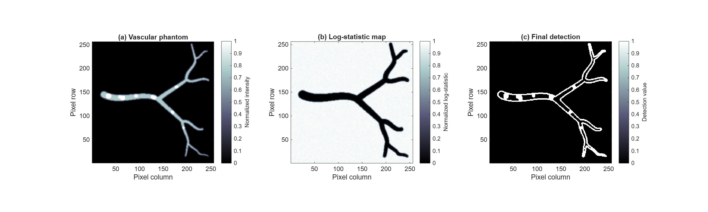
Fig. 1. (a) Simulated vascular phantom containing spatially distributed contrast-agent inclusions represented by bright regions. Contrast-agent visibility was intentionally enhanced for visualization purposes and does not represent a quantitative physical scale. (b) Accumulated log-statistic map obtained from noisy photoacoustic realizations, showing clear separation between signal-containing regions and the noisy background after cumulative evidence integration. (c) Final detection result produced by the proposed dual-wavelength spectroscopic framework
Photoacoustic imaging is a hybrid biomedical imaging modality that combines optical excitation with ultrasonic detection, enabling spectroscopic discrimination of chromophores embedded within biological tissue. One of the principal challenges in spectroscopic photoacoustic imaging is the reliable detection of exogenous contrast agents in the presence of endogenous absorbers such as deoxyhemoglobin (Hb) and oxyhemoglobin (HbO$_2$), particularly under noisy acquisition conditions and spectral overlap.
In this work, we propose a dual-wavelength spectroscopic framework for robust contrast-agent detection without explicitly estimating chromophore concentrations. Rather than solving a spectral unmixing problem, the proposed framework determines whether a target contrast agent is present or absent at a given spatial location based on its wavelength-dependent photoacoustic response.
The proposed framework begins with an optimal wavelength-pair selection criterion based on a max–min spectral separation index. For a target chromophore $m$ and interferent $n$, the spectral separation index is defined as
$$ S_m,n(\lambda^+,\lambda^-)= \frac{ǎrepsilon_m(\lambda^+)}{ǎrepsilon_n(\lambda^+)} ḑot \frac{ǎrepsilon_n(\lambda^-)}{ǎrepsilon_m(\lambda^-)}. $$
where $\varepsilon(\lambda)$ denotes the absorption spectrum of the corresponding chromophore. This criterion identifies wavelength pairs that maximize spectral contrast reversal between the target absorber and endogenous interferents. The optimal wavelength pair is obtained by maximizing the following criterion:
$$ J_m(\lambda^+,\lambda^-) = \min_n S_m,n(\lambda^+,\lambda^-) $$
over all feasible wavelength pairs satisfying spectral inequality constraints that favor the target chromophore over endogenous interferents at $\lambda^{+}$ while reversing this relationship at $\lambda^{-}$.
The proposed methodology was evaluated through photoacoustic simulations using deoxyhemoglobin (Hb) and oxyhemoglobin (HbO$_2$) as endogenous interferents and glycol chitosan-coated gold nanoparticles as the target absorber. Curved vascular phantoms containing spatially distributed inclusions were generated, and additive Gaussian noise was incorporated into the simulated photoacoustic signals. A cumulative log-regularized statistic derived from repeated noisy realizations of a multiplicative spectral ratio was introduced to accumulate spectroscopic evidence while improving robustness against noise and spectral fluctuations. Representative simulation and detection results are shown in Figure 1.
Preliminary results demonstrate consistent detection of gold nanoparticle inclusions under noisy conditions, a significant reduction in false positives through signal-derived support constraints, and improved detection stability through cumulative spectroscopic evidence integration. These results establish the proposed dual-wavelength spectroscopic framework as a proof of concept for robust contrast-agent detection in photoacoustic imaging. Future work will focus on validating the proposed framework under increasingly realistic photoacoustic imaging conditions and benchmarking its performance against conventional spectral unmixing methods.